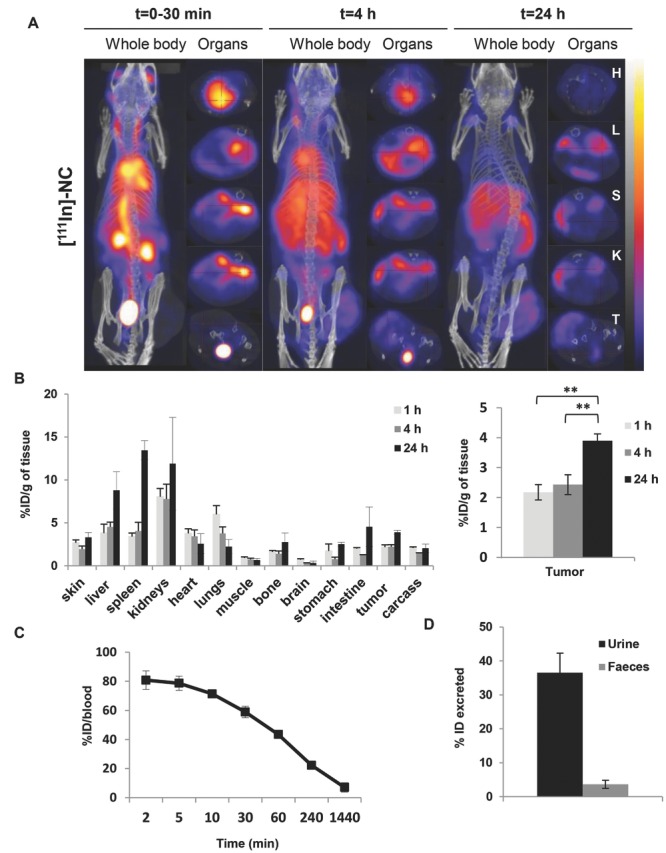
Figure 7.

In vivo whole body 3D SPECT/CT imaging and biodistribution of NC-111In in CT26 tumor-bearing balb/c. Mice were i.v injected with NC-111In at a dose of 600 mg polymer per kg. Mice for SPECT/CT imaging with tumor inoculated at one side only while tumor inoculated bifocally for gamma scintigraphy studies. A) Whole body 3D SPECT/CT imaging at 0–30 min, 4 and 24 h post-injection with scanning time of 40–60 min each. Cross-sections were from heart (H), liver (L), spleen (S), kidney (KI), and tumor (T) at equivalent time points. Tumor accumulation was observed at 4 h post-injection and was enhanced over time. B) Organ biodistribution profile values were expressed as percentage injected dose per gram tissues (% ID/g) at 1, 4, and 24 h after injection of 600 mg PLGA-PEG/kg. Inset in A is showing the uptake in tumors. Blood clearance profile is shown in C); excretion profile at 24 h is shown in D). Signals were quantified by gamma scintigraphy. Results are expressed as means ± SD (n = 3–4). *P < 0.05, **P < 0.01 (one-way ANOVA test).
