
Fig. 2.
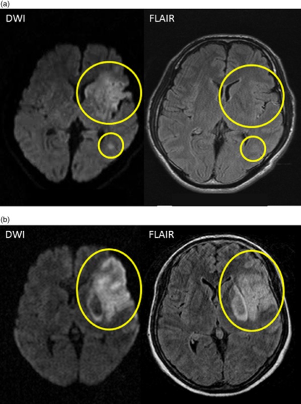
Examples of magnetic resonance imaging (MRI) inclusion and exclusion criteria. (a) A negative fluid-attenuated inversion recovery (FLAIR) pattern shows an acute ischemic lesion clearly visible on diffusion-weighted imaging (DWI), but no marked parenchymal hyperintensity visible on fluid-attenuated inversion recovery (FLAIR) corresponding to the DWI lesion (yellow circles). (b) A positive FLAIR pattern shows an acute ischemic lesion clearly visible on DWI and clear parenchymal hyperintensity on FLAIR corresponding to the acute DWI lesion (yellow circle).