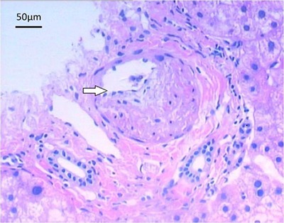
Figure 4. Microscopic image of a section of the liver taken following biopsy. The arrow shows the enhanced thickness of the hepatic portal vein wall in the portal area. Note that the venous lumen is not fully occluded. Hematoxylin-eosin staining, 100×.

