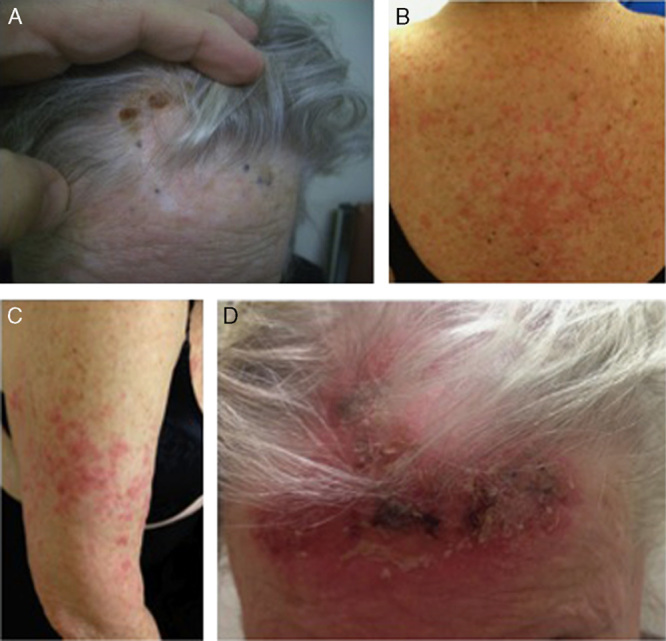
FIGURE 2.

Patient 2’s response. A, Primary lesions are nine 1–3 mm gray to purple-black macules and thin papules on the frontal scalp. The crusted brown lesions were the sites of biopsy. B and C, Following 1 sensitization dose of Bacillus Calmette-Guérin, the patient developed numerous edematous, pink-red papules and plaques on the back and proximal bilateral arms, consistent with lichenoid drug eruption. D, Following topical imiquimod application, most lesions faded and developed inflammatory changes and hemorrhagic crusting, and 6 weeks after discontinuation of therapy, signs of complete regression were observed (images not shown).
