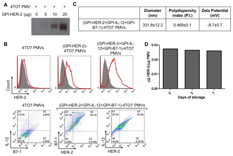
Figure 3. Protein transfer of GPI-HER-2 with GPI-ISMs onto PMVs derived from 4TO7 tumor tissue.
(A) Protein transfer mediated incorporation of GPI-HER-2 onto 4TO7 PMVs is concentration dependent. 20 μg of 4TO7 PMVs were protein transferred with increasing concentrations of GPI-HER-2. Incorporation of GPI-HER-2 was analyzed by SDS PAGE and western blot analysis. (B) Flow cytometry analysis of 4TO7 PMVs protein transferred with GPI-HER-2 +/− GPI-ISMs. 4TO7 PMVs were protein transferred with GPI-HER-2 along with GPI-IL-12 and/or GPI-B7-1. Then PMVs were stained with fluorescein conjugated mAbs and analyzed by flow cytometry. (C) Protein transfer of GPI-APs onto 4TO7 PMVs does not affect PMV size. The diameter, polydispersity index and zeta potential of unmodified or protein transferred 4TO7 PMVs were analyzed using a Zetasizer. (D) HER-2 incorporated onto 4TO7 PMVs by protein transfer remains stably expressed at room temperature. 4TO7 PMVs modified by protein transfer with GPI-HER-2 were stored at room temperature. Each time point, PMVs were washed by centrifugation with PBS and the presence of HER-2 was determined by SDS PAGE and western blot analysis by staining against HER-2.

