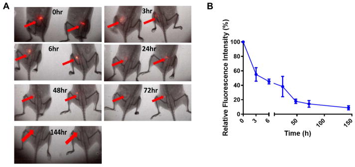
Figure 8. Vaccination with H-PMVs leads to prolonged HER-2 persistence at the vaccination site.
Purified GPI-HER-2 was fluorescently labeled with IRDye 800CW and then protein transferred onto 4TO7 PMVs. BALB/c mice (n = 4) were injected s.c. on the hind flank with 25 μg of the resulting tagged H-PMVs. (A) Optical imaging was conducted using the Kodak In vivo FX imaging system (Carestream Health Inc.) at the indicated time points (ex: 720 nm; em: 790 nm). Representative images of mice are shown. (B) Image J analysis was used to quantify the average fluorescence intensity at the injection sites. Regions of Interests were selected for measuring the mean fluorescence intensity (MFI) of the protein at the vaccination site and corresponding body background. Signal to body background (S/B) ratio was calculated from the MFI of the injection area divided by the MFI of the body background area. Data shown is the mean S/B ratio ± standard error from three to four mice in which n = 4 was used for the initial time points and n = 3 was used for the 24 h time point onwards.

