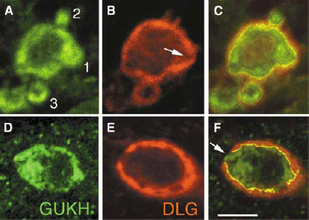
Figure 3. GUKH and DLG Are Dynamically Expressed during Bouton Budding.
(A–C) First instar NMJs showing three budding boutons (“1,” “2,” and “3”), double labeled with (A and D) anti-GUKH and (B and E) anti-DLG. (C) and (F) are merged images from (A) and (B) and from (D) and (E), respectively. Numbers 1–3 in (A) indicate stages of bouton budding (1, protrusion; 2, bud separation; 3, new bouton formation). Arrow in (B) and (F) points to low DLG levels at the site of bouton protrusion. Scale bar represents 1.5 µm in (A)–(C) and 3 µm in (D)–(E).

