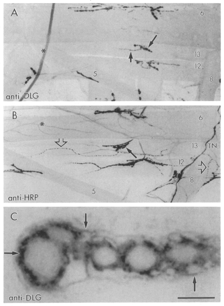
Figure 1. Confocal Micrographs of dlg- and HRP-Like Immunoreactivity at the Body Wall Muscles of Wild-Type Drosophila Larvae.
(A) Type Ib synaptic boutons stain with anti-dlg antibodies (long arrow). Type Is boutons also stain (short arrow), but to a lesser degree.
(B) Neuromuscular junctions stained with anti-HRP antibodies, which stain all bouton types. Note the presence of type II boutons (open arrows) in a subset of muscles. Long arrow points to a type Ib bouton in muscle 13. Asterisk marks a branching trachiole.
(C) High magnification view of dlg-like immunoreactivity in a string of type Ib boutons at muscle 6. Focus was at the midlevel of the boutons. Arrows point to the perisynaptic network. TN, transverse nerve. Scale bar equals 90 µm in (A) and (B), 5 µm in (C).

