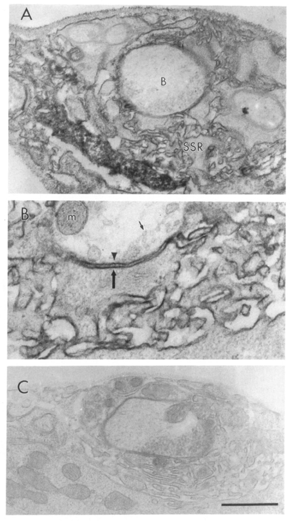
Figure 2. Immunolocalization of dlg at Type Ib Boutons.
(A) Type Ib boutons (indicated with a B) at muscle 6 showing immunoreactivity associated with the SSR. (B) High magnification view of the presynaptic (arrowhead) and postsynaptic (large arrow) membrane, and the associated immunoreactivity. Note that immunoreactivity is concentrated at the SSR, and to a lesser extent, to the presynaptic bouton border. Small arrow indicates a synaptic vesicle. Immunoreactivity was visualized using the peroxidase reaction with DAB as a substrate in samples fixed with paraformaldehyde and 0.01% glutaraldehyde. m, mitochondria. (C) Type Ib bouton from a control sample in which the primary antibody was omitted. Scale bar equals 1 µm in (A) and (C), 0.43 µm in (B).

