FIGURE 5.
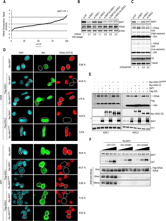
ASXM of ASXL1/2 stimulates BAP1 DUB activity. A, siRNA screen for DUBs that coordinate H2Aub levels. Following transfection with siRNA DUB library, HeLa cells were fixed and immunostained for H2Aub Lys-119 (H2Aub). The fluorescence signal was determined, and the values were used to derive the Z scores. B, knockdown of BAP1 or concomitant Knockdown of ASXL1 and ASXL2 induces a significant increase of the global level of H2Aub. U2OS cells were transfected with siRNA as indicated and harvested 4 days post-transfection for immunoblotting using the indicated antibodies. Quantification of band intensity for H2Aub was conducted relative to the non-target siRNA control (siNT). C, increase of H2Aub levels following BAP1 depletion is not due to a global increase of H2A. U2OS cells were transfected with siRNA of BAP1, ASXL1, and ASXL2 and harvested, 4 days post-transfection, for immunoblotting using the respective antibodies. Tubulin was used a loading control for soluble proteins and histone H3 as a loading control for histones levels. Quantification of band intensity for H2Aub was conducted relative to the non-modified histone H2, and the values were then normalized to the non-target siRNA control (siNT). D, ASXL1/2 promote BAP1 DUB activity toward H2Aub in vivo. U2OS cells (top panel) or 293T cells (bottom panel) were transfected with either Myc-BAP1 (0.5 μg) or Myc-BAP1 C91S (0.5 μg) expression constructs in the presence or absence of FLAG-ASXL1/2 (4 μg) expression constructs. Three days post-transfection, cells were harvested for immunostaining using the indicated antibodies. The cells overexpressing BAP1 and BAP1C91S were encircled. Note that the transfections were conducted with plasmid ratios optimized to ensure that most BAP1-transfected cells also express ASXL1 or ASXL2. Cells overexpressing BAP1 were counted for change in H2Aub signal. The percentages at the right of the panel represent the number of cells showing very low signal of H2Aub versus the total number of BAP1-expressing cells. E, 293T cells were transfected as indicated using FLAG H2A (0.2 μg), BAP1 (1 μg), Myc-ASXL1 (4 μg), or Myc-ASXL1 ΔASXM (4 μg) vectors (left panel) and Myc-ASXL2 (4 μg) or Myc-ASXL2 ΔASXM (6 μg) vectors (right panel) and harvested, 3 days later, for immunoblotting. F, in vitro DUB assay of nucleosomal H2A using recombinant His-BAP1 (8 ng, 2 pm) in the presence of increasing amounts of recombinant GST-CTD, GST-ASXM1, or GST-ASXM2 (0.6, 1.2, 2, and 4 pm). β-Actin, tubulin, or YY1 was used as loading controls.

