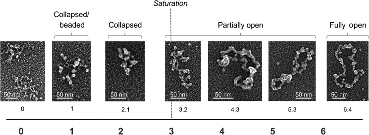
FIGURE 2.
Electron microscopy of human wild-type mtSSB protein bound to the M13 DNA template. The binding reaction was performed at 30 mm KCl and 4 mm MgCl2. The numbers below individual images indicate the ratio of SSB tetramers per 100 nucleotides of template DNA. The dashed line indicates the concentration of SSB tetramers that would result in saturation of the template DNA molecules (as predicted from the ssDNA binding site size of 35 nt/tetramer). The following species were distinguished: collapsed/beaded, collapsed, partially open, and fully open.

