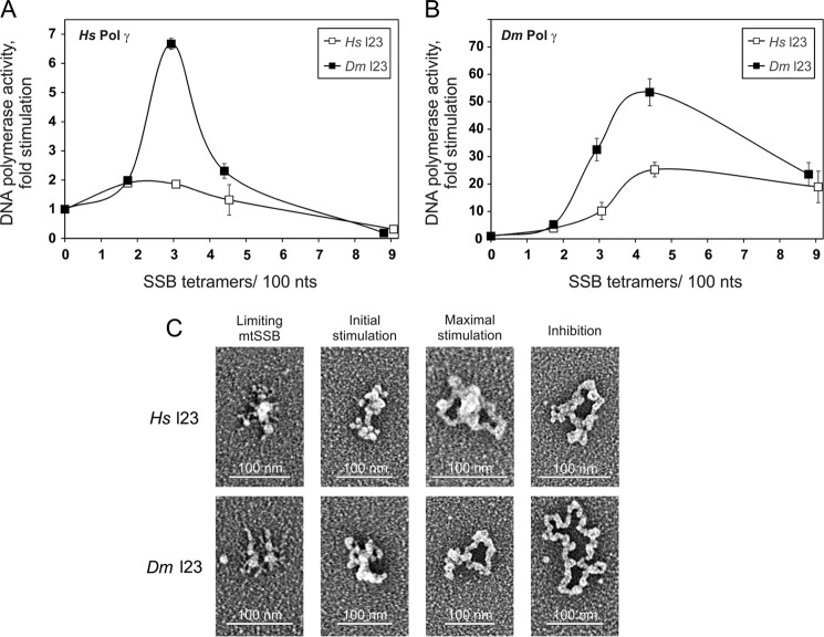
FIGURE 4.
Stimulation of the DNA polymerase activity of Pol γ and template DNA organization by loop 2,3 variants of HsmtSSB and DmmtSSB. A and B, a DNA polymerase assay was performed as described under “Experimental Procedures,” under conditions as described in the legend to Fig. 3A, except that the loop 2,3 mtSSB variants (H. sapiens (open squares) or D. melanogaster (closed squares)) were used. C, electron microscopy of H. sapiens (top) and D. melanogaster (bottom) loop 2,3 variants of mtSSB on M13 DNA. The binding reaction was performed at 30 mm KCl and 4 mm MgCl2. The images are representative of template species formed at the following ratios of SSB tetramers/100 nucleotides, which correspond to the indicated individual phases of the stimulation of HsPol γ activity: limiting mtSSB, 1.7 Hsl2,3 mtSSB and 1.2 Dml2,3 mtSSB; initial stimulation, 3.4 Hsl2,3 mtSSB and 1.8 Dml2,3 mtSSB; maximal stimulation, 4 Hsl2,3 mtSSB and 2.5 Dml2,3 mtSSB; inhibition, 6.8 Hsl2,3 mtSSB and 7.2 Dml2,3 mtSSB (also see Table 1). Error bars, S.D.

