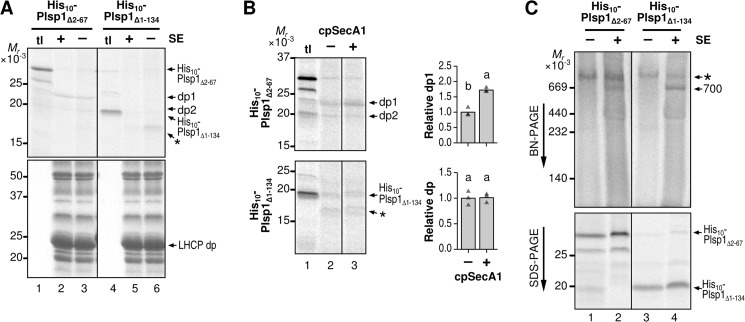
FIGURE 8.
Requirements of TMD within Plsp1 for spontaneous membrane insertion, cpSecA1-dependent transport, and association with the stroma complex. A, effects of SE on transport of His-tagged Plsp1 variants. Radiolabeled proteins indicated on top were incubated with isolated chloroplast membranes with 5 mm ATP in the light for 30 min. After treatment with thermolysin, products were separated by SDS-PAGE. Proteins on the same gel were visualized by phosphorimaging (top panel) or Coomassie Brilliant Blue staining (bottom panel). tl lanes were loaded with 10% of translation products used for the assay. B, effects of cpSecA1 on transport of His-tagged Plsp1 variants. Radiolabeled proteins indicated at left were incubated with isolated chloroplast membranes in the light in the absence of SE, without (−) or with (+) 0.375 μg of recombinant cpSecA1, followed by thermolysin treatment. Products were separated by SDS-PAGE and visualized using phosphorimaging. Shown at right are quantifications of data from three repetitions (triangles) and their means (bars) with letters indicating Tukey HSD groupings above for α = 0.05. C, incorporation of His-tagged Plsp1 variants into the 700-kDa complex in SE. Radiolabeled proteins indicated on top were incubated without (−) or with (+) SE for 30 min at room temperature, followed by transfer to ice and addition of cold BN-PAGE sample buffer. Samples were then separated by BN-PAGE (top) or SDS-PAGE (bottom) and visualized using phosphorimaging. The 700-kDa complex incorporating the radiolabeled Plsp1 variants (700) and a larger complex present in the translation product mixture (*) are indicated to the right of the BN-PAGE gel. The bands corresponding to each of the two Plsp1 variants are indicated at the right of the SDS-PAGE gel.

