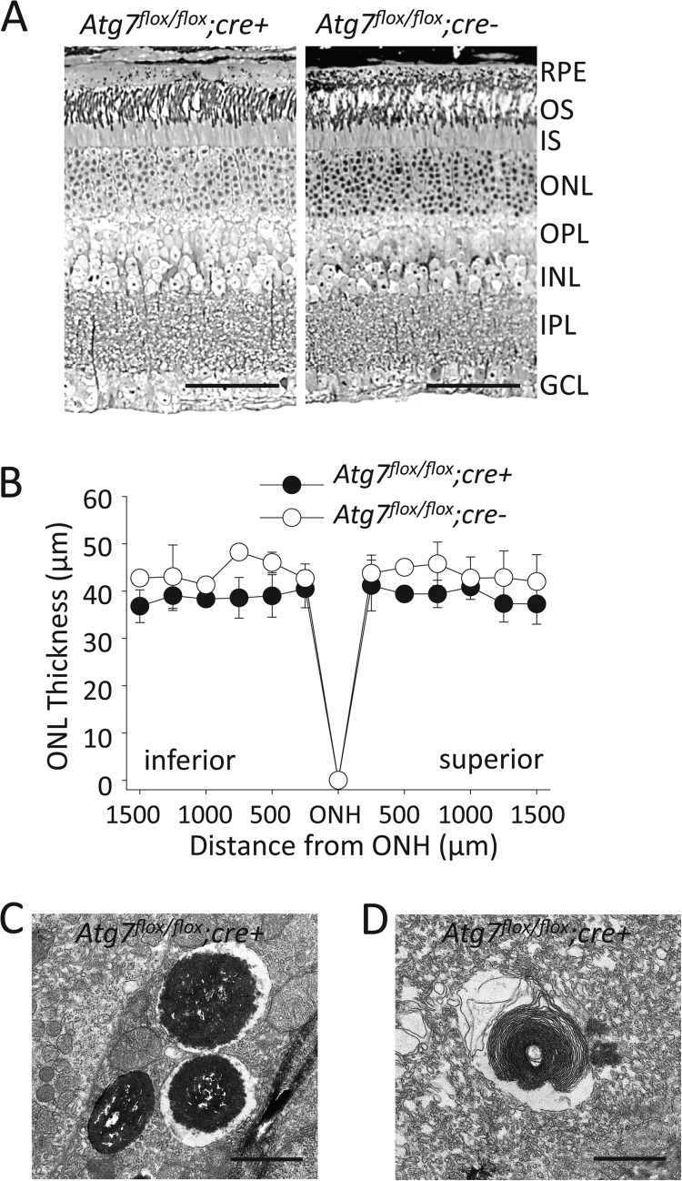
FIGURE 5.
Loss of Atg7 in the RPE does not lead to age-associated retinal disease A, toluene blue staining of retinal sections from 12-month-old Atg7flox/flox;cre+ and Atg7flox/flox;cre- mice. OS, outer segment; IS, inner segment; ONL, outer nuclear layer; OPL, outer plexiform layer; INL, inner nuclear layer; IPL, inner plexiform layer; GCL, ganglion cell layer. Scale bar 50 μm. B, outer nuclear layer (ONL) thickness was measured on DAPI stained retinal sections from 12-month-old Atg7flox/flox;cre+ and Atg7flox/flox;cre- mice, n = 4 animals per group. Error bars indicate the mean ± S.D. ONH, optic nerve head. C and D, electron microscopic images displaying dysfunctional autophagy in the RPE of Atg7flox/flox;cre+ mice, including partial digestion of melanosomes (C) and rod outer segments (D). Scale bar 1 μm.

