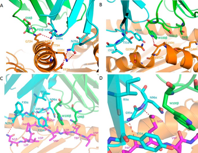
FIGURE 7.
Interactions of TCR C7 with HLA-A2 and the NLV peptide. A, interactions of CDR1α, CDR3α, and CDR2β with the HLA-A2 α1 helix. The side chains of contacting residues are drawn in stick representation with carbon atoms in cyan (CDR1α and CDR3α), green (CDR2β), or orange (HLA-A2), nitrogen atoms in blue, and oxygen atoms in red. Hydrogen bonds are indicated by red dashed lines. B, interactions of CDR1α, CDR2α, and CDR3β with the HLA-A2 α2 helix. The side chains of contacting residues are drawn with carbon atoms in cyan (CDR1α and CDR2α), green (CDR3β), or orange (HLA-A2). C, interactions of CDR1α, CDR3α, and CDR3β with the NLV peptide. The side chains of contacting residues are drawn with carbon atoms in cyan (CDR1α and CDR3α), green (CDR3β), or magenta (NLV). D, close-up of interactions between C7 and P5 Met.

