
Fig. 2.
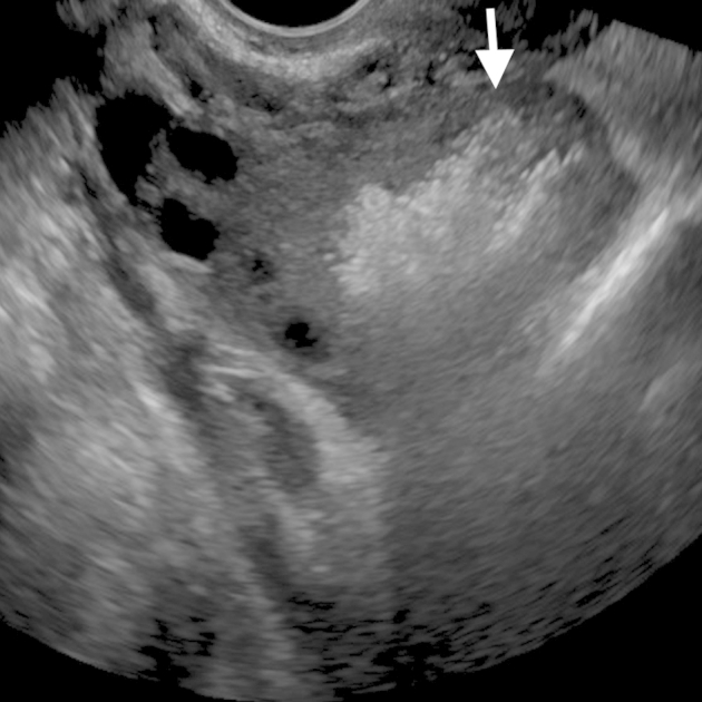
Initial pelvic ultrasound obtained within 3 hours of the computed tomography shows an echogenic mass in the right ovary with “dirty” posterior acoustic shadowing (annotated by the white arrow).
Official websites use .gov
A
.gov website belongs to an official
government organization in the United States.
Secure .gov websites use HTTPS
A lock (
) or https:// means you've safely
connected to the .gov website. Share sensitive
information only on official, secure websites.
Initial pelvic ultrasound obtained within 3 hours of the computed tomography shows an echogenic mass in the right ovary with “dirty” posterior acoustic shadowing (annotated by the white arrow).