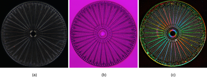
Figure 2. Images of a diatom Arachnoidiscus.
All images were taken in white light. The picture size is 190 μm × 190 μm. The maximum retardance is 5 nm. The left photo (a) depicts a case with crossed linear polarizer and analyzer. The middle image (b) illustrates a case with full-wave plate inserted into the optical path. The right picture (c) shows polarization polychromatic image with the brightness corresponding to retardance and hue representing the principal (birefringent) axis orientation.

