
Fig. 5.
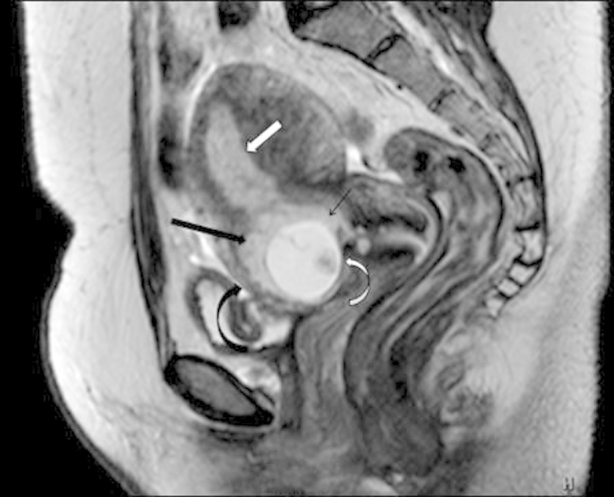
Sagittal T2-weighted imaging showing a gestational sac (curved white arrow) implanted within the anterior myometrium of the lower uterine segment in the region of the scar of previous cesarean section. The gestational sac shows a well-formed fetal pole within and is surrounded by a well-appreciated decidual reaction (thick black arrow). Anterior myometrium anterior to the gestational sac (curved black arrow) is thinned out. Posteriorly, the gestational sac is seen extending into the endometrial cavity in the lower uterine segment (thin black arrow). The posterior myometrium shows good wall thickness.