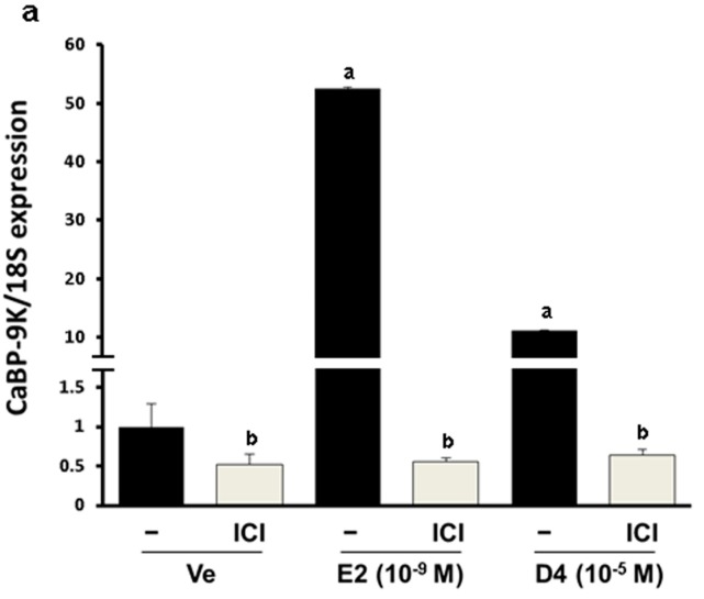
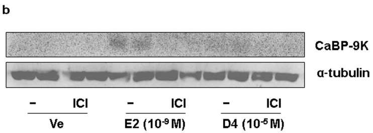
Figure 1.
CaBP-9K expression in GH3 cells at the mRNA (a); and protein (b) levels. Results presented in the bar graph are divided according to chemical administered and subdivided according to the presence or absence of ICI 182 780. Ve, vehicle; E2, 17β-estradiol; D4, octamethylcyclotetrasiloxane. a p < 0.05 vs. vehicle; b p < 0.05 vs. without ICI. Data are presented as the mean ± SD.


