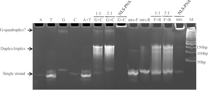
Figure 2. Assessment of assembly of oligonucleotide scaffolds and binding to the complementary PNA.

Single-strand oligomers (lanes 1–4), mixed sequences (lanes 9 and 10), the oligomer 12A-5A plus 12T-5A (lane A + T), the oligomer 12G-5A plus 12C-5A (lane G + C, ratio of 1:1 or 2:1), or mixed-sequences forward plus reverse (lane F + R, ratio of 1:1 or 2:1) were annealed in a total volume of 20 μL and analyzed by polyacrylamide gel electrophoresis. Lanes 8 and 13 represent the equimolar complementary NLS-PNA (final concentration of 2.5 μM) annealed with the oligonucleotide scaffold. The bands corresponding to single strand, double/triplex strand, or G-quadruplex are indicated (arrows).
