Figure 5.
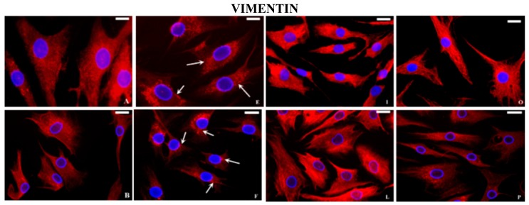
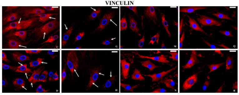
Indirect immunofluorescence microscopy. Basal conditions: the vimentin filaments in normal chondrocytes (A) were organized in all the cytoplasm as a network, crossing from the cell periphery to the nuclear membrane; in OA cells (B) the distribution is partially altered. Normal (C) and OA chondrocytes (D) incubated with anti-vinculin antibody show a punctate pattern under the plasma membrane (arrows). Incubation with IL-1β: normal and OA chondrocytes show a reduced fluorescence and a destruction of both vimentin (E, F respectively, arrows) and vinculin (G, H respectively, arrows) filaments. Cyclical hydrostatic pressure (HP): fluorescent signal of vimentin and vinculin proteins in normal (I, M respectively) and OA chondrocytes (L, N respectively). Exposure to HP + IL-1β: fluorescent signal of vimentin and vinculin proteins in normal (O, Q respectively) and OA chondrocytes (P, R respectively). Nuclei (blue) were stained with DAPI and anti-vimentin and anti-vinculin (red). Bar: 50 μm.


