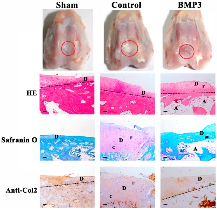
Figure 2.
Macroscopic and histological observation of the repaired tissue in full-thickness defect at eight weeks post-operation. The red circle regions are the defects of articular cartilage. Sections were stained with HE, Safranin O, or immunohistochemical collagen type II (Anti-Col2). Sham, sham-operated group; Control, group only treated with collagen membrane without BMP3 as negative control; BMP3, group treated with the collagen membrane and BMP3 at 100 ng/cm2, respectively. The dash line stands for the boundary between articular cartilage and subchondral bone. D, C, F, A stand for defect area, chondrocyte-like cells, fibrocartilage cells, cavities, respectively. Scale bar is 100 μm.

