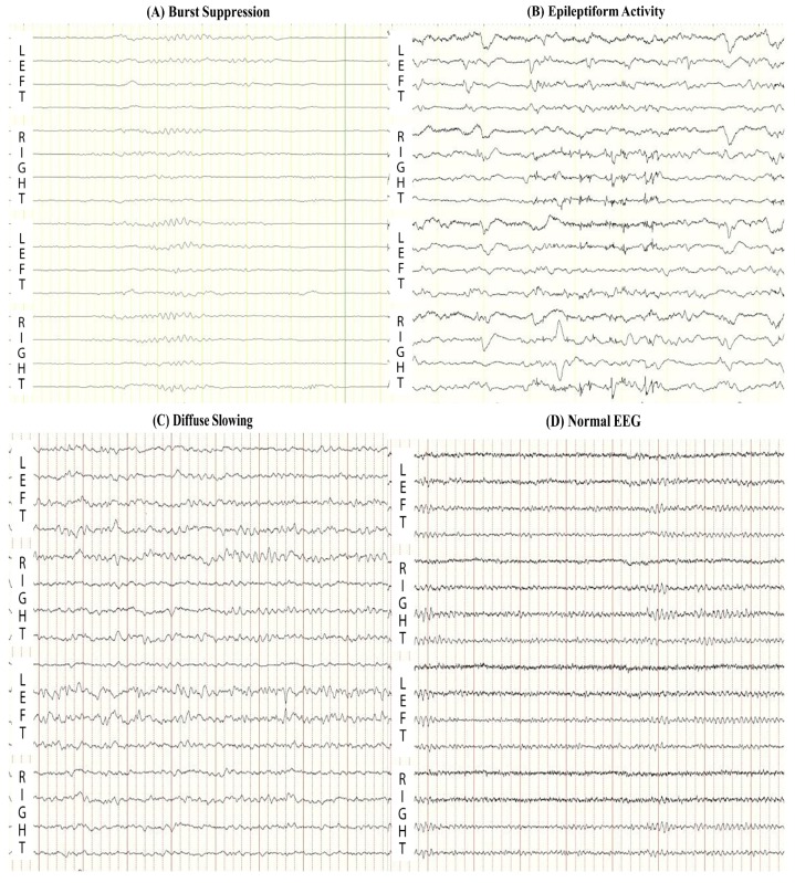
Figure 1.
Representative abnormal electroencephalography (EEG) patterns: (A) Burst Suppression (the presence of bursts with amplitudes higher than 20 μV, followed by the intervals of at least 1 s with suppression of EEG activity less than 20 μV) and (B) Epileptiform Activity (including seizures and generalized periodic discharges) are associated with poor outcome; (C) Continuous Diffuse Slowing (EEG activity with a dominant frequency less than 8 Hz) and (D) Normal EEG at 12-h after resuscitation are associated with good outcome.

