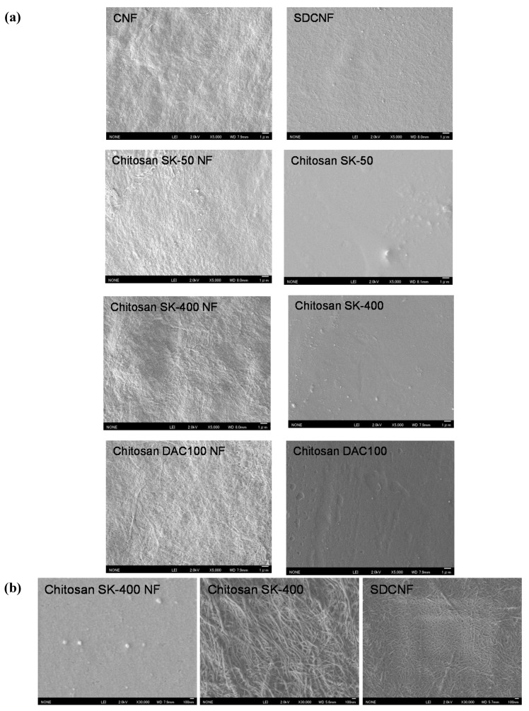
Figure 1.
Field emission scanning electron microscopic (FE-SEM) micrographs of sheets. (a) SEM micrographs of chitin nanofiber (CNF), surface-deacetylated chitin nanofiber (SDCNF), chitosan NFs and chitosan sheets with low magnification (×5000); (b) Representative surface image of chitosan SK-400 NF, Chitosan SK400, and SDCNF sheets with high magnification (×30,000).

