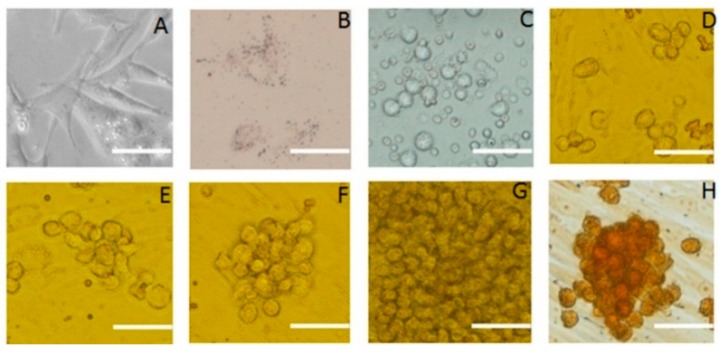
Figure 1.
Morphology of SSCs and Sertoli cells in culture. Appearance under the microscope and various staining properties were used to identify the cells. (A) Sertoli cells derived from porcine testicular tissue; (B) Oil red-o staining of Sertoli cells; (C) Primary SSCs derived from porcine testicular tissue; (D–G) Colony of SSCs grown on feeder cells for 3, 5, 7, 12 days; (H) SSCs clone cells stained for alkaline phosphatase activity; the image indicates strong alkaline phosphatase activity. Scale bar: 5 μm.

