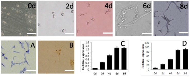
Figure 5.
Morphology and gene expression of SSCs differentiated into neuron-like cells in vitro. Appearance under the microscope and various staining properties were used to confirm their identity.Schematic represents the strategy used for in vitro differentiation. 0d, 2d, 4d, 6d, 8d mean the same, 2th, 4th, 6th, 8th day after induction. (A) Toluidine blue staining of SSCs-derived cells; (B) Immunocytochemcal staining of SSCs-derived cells; (C) Expression of Nestin and (D) β-tubulin, both measured by mRNA levels during induction. Scale bar: 5 μm.

