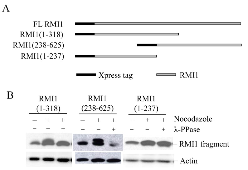
Figure 3.
Mapping RMI1 mitotic phosphorylation sites. (A) Schematic representation of full-length (FL) Xpress-RMI1 and truncated fragments; (B) 293T cells were transfected with the constructs shown above. Thirty-two hours later, cells were treated with nocodazole or not for 16 h prior to harvest. Extracted proteins were treated with λ-phosphatase or not as shown, then resolved by 8% polyacrylamide gel and blotted with anti-Xpress antibody.

