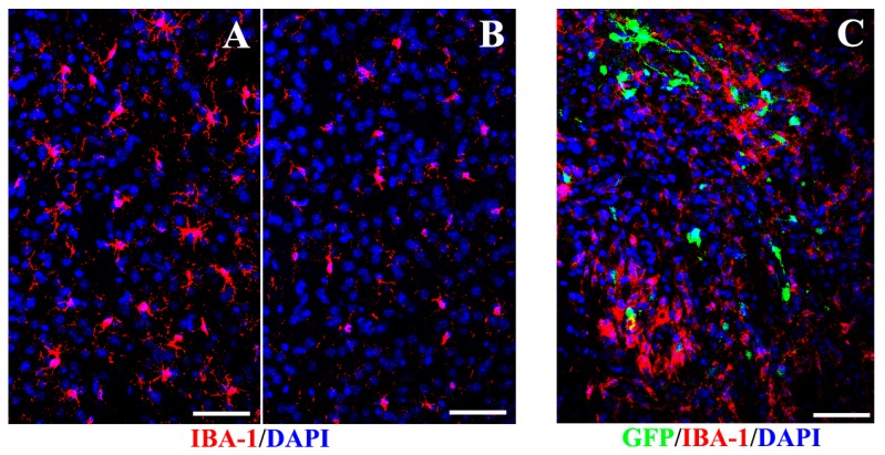
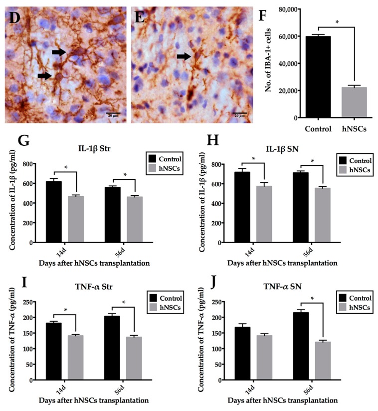
Figure 9.
Effects of hNSCs-induced inhibition of host microglia and proinflammatory cytokines. Fewer IBA-1 positive microglia (red) presented in the striatum of hNSCs-treated mice (B) than the control animals (A); (C) Transplanted hNSCs (green) gathered local microglia with activated forms (red) to a limited area around the xenografts. Cell nuclei were counterstained with DAPI (blue) (A–C). Most microglia presented fully activated forms in control mice (arrows in D), while arrow in (E) indicated the ramified microglia (resting state of microglia) in transplanted animals. Cell nuclei were hematoxylin counterstained in (D,E). The numbers of microglia in different groups were compared as illustrated in (F). Additionally, the expression of IL-1β (G,H) and TNF-α (I,J) was downregulated in the striatum and SN of mice receiving hNSCs compared with the control animals. Data are expressed as mean ± SEM (* p < 0.05; two-tailed Student’s t-test). Scale bars represent 50 μm in (A–C); 20 μm in (D,E). IBA-1, ionized calcium-binding adapter molecule 1; IL-1β, interleukin-1β; TNF-α, tumor necrosis factor-α.


