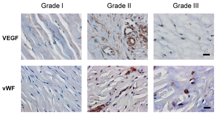
Figure 5.
VEGF expression and vessel density in tissue samples of retinaculum were dependent upon disease severity. Serial sections of three grades (I–III) were stained with vascular endothelial growth factor (VEGF) and Von Willebrand’s factor (vWF) antibodies. More blood vessels were distributed in the grade II and grade III tissues of retinaculum (immunohistochemical staining, 400× magnification, scale bar = 10 μm).

