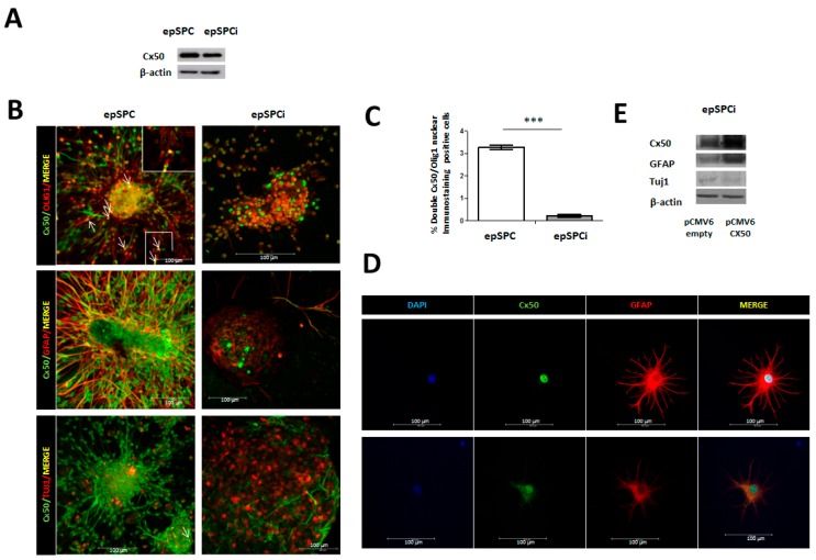
Figure 1.
Cx50 expression in epSPC compared to epSPCi and their derived differentiated cells. (A) Cx50 exhibited higher protein expression in epSPC compared to activated epSPCi; (B) Immunocytochemical analysis of Cx50 simultaneously with the markers for the neuronal lineage (GFAP for astrocytes, Olig1 for oligodendrocytes and Tuj1 for neurons) in epSPC and epSPCi under spontaneous differentiation conditions for 24 h. The inset shows a zoom in area for easier visualization of Olig1 and Cx50 co-localization. Arrows point double-stainings. Scale bar = 100 µm; (C) Quantification of co-localization for Cx50 and Olig1; *** p < 0.001 vs. epSPC; (D) Immunocytochemistry of Cx50 and GFAP antibodies in primary culture of mature astrocytes isolated from non-injured spinal cord rats. Scale bar = 100 µm; (E) Modulation of GFAP and Tuj1 in epSPCi after over-expression of Cx50 by using pCMV6-Cx50 expression vector in comparison to pCMV6-empty (control).

