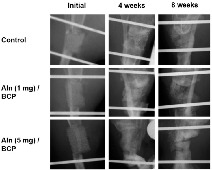
Figure 6.
Plain radiographs of rat tibial defect model. The sharp margin of the osteotomy sites were disappeared with laps of time at four and eight weeks in all specimens. More bone formation and high radio-opaque consolidation of the defect areas were observed at eight weeks in Aln (5 mg)/BCP scaffold model specimens. However, no solid bony bridging was observed in any of the all three groups, the Aln/BCP groups showed relatively abundant callus formations compared to the control group.

