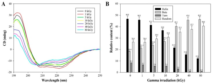
Figure 5.
Change in the secondary structure of AtTDX by γ-irradiation. (A) Far-UV CD spectra of non-irradiated or irradiated AtTDX proteins in 10 mM Tris-HCl (pH 7.4) buffer. The ellipticity of the CD spectra was expressed in millidegrees (mdeg); (B) The comparison of the secondary structure index values (%) was based on the far UV-CD spectra results of AtTDX under different γ-irradiation doses. Data are the means of at least three independent experiments.

