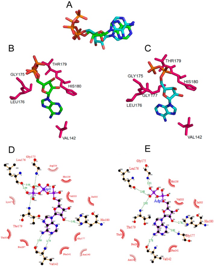
Figure 1.
The amino acid residues forming the nucleotide-binding pocket of DnaA protein bound to ADP and ATP adopt different confirmations. (A) Overlap of the conformations of ADP and ATP present in the modeled E. coli DnaA protein in their bound state; (B,C) Amino acid residues interacting with the ATP and ADP. Figures were generated using PYMOL software; (D,E) Molecular interactions of residues in the nucleotide binding pocket with bound ATP (D) or ADP (E), hydrogen bond interactions are indicated as dashed lines in green and the van der Waals interaction are indicated as half-circles in red wire diagrams. The numbers indicate the hydrogen bonding distance between the atoms in Å units. The Figures were generated using PDBSum.

