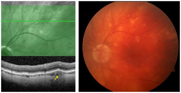
Figure 1.
(Right) Color fundus photograph of a 59-year-old male patient with multifocal choroiditis secondary to sarcoidosis demonstrates multiple round, creamy, subretinal lesions in the superotemporal quadrant of the left eye; (Left) optical coherence tomography study through the lesion showed that the lesion primarily involves the choroid (arrow).

