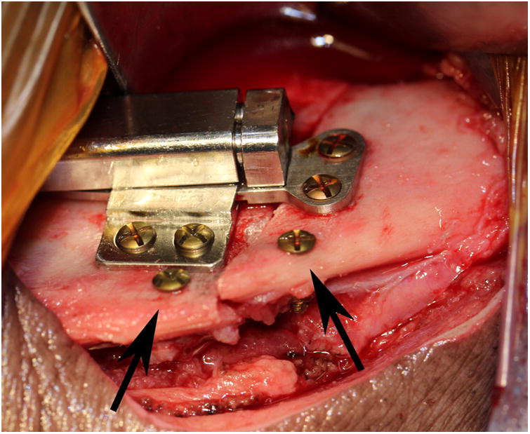
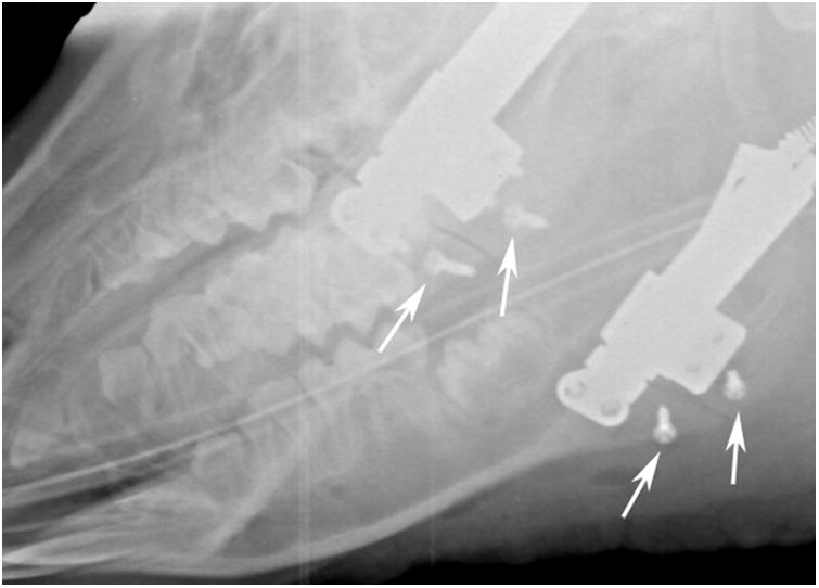
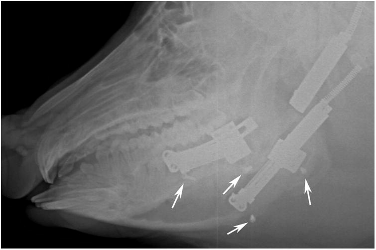
Figure 1.


A, Intraoperative photograph of the device fixed across the osteotomy with markers screws (arrows) placed at the inferior border. B, Immediate postoperative radiograph showing bilateral osteotomies with marker screws (white arrows) C, End-Fixation radiograph showing the lengthening of the mandible with increased distance between marker screws (white arrows). The right side distractor became unattached during the fixation period with some loss of lengthening. The left side distractor is fully extended. The regenerate appears radiodense for both sides (scores of 3).
