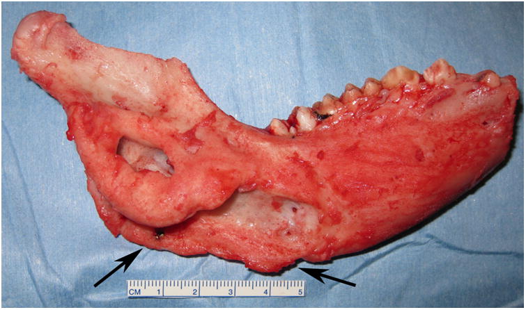
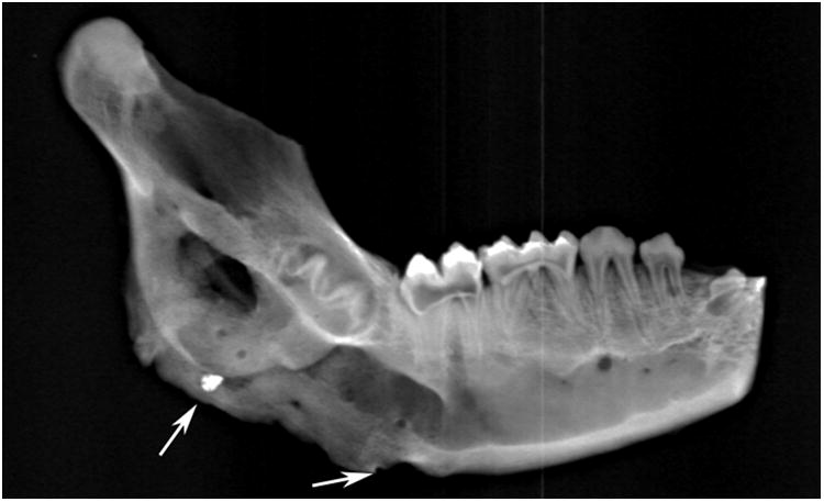
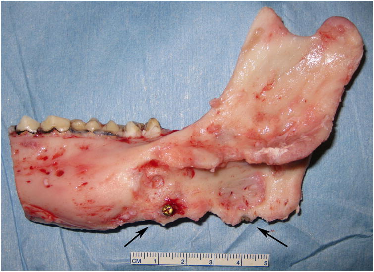
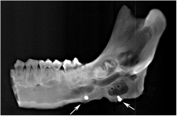
Figure 2.


Right side hemimandible photograph (A) and radiograph (B) showing strong clinical bone fill (score of 3) and radiodensity (score of 3) within the distraction gap. The left side hemimandible photograph (C) and radiograph (D) showed similar results (score of 3 for each). Position of marker screws marked with black arrows.
