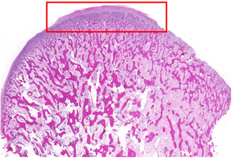
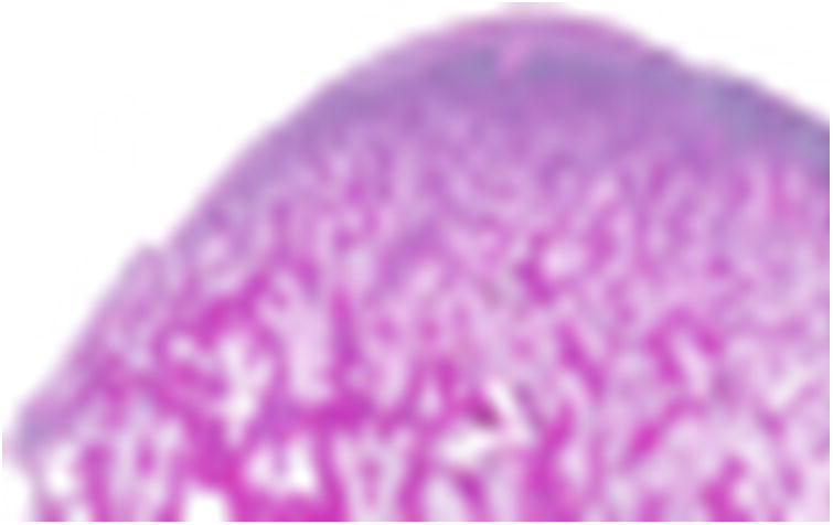
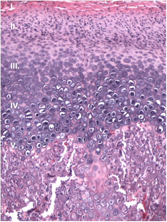
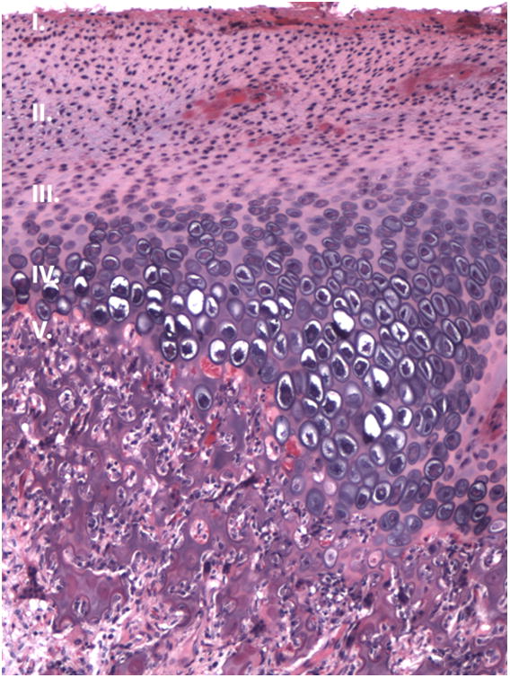
Figure 4.


Condyle histology. (H&E, sagittal view). A-B, Photomicrograph of the (A) right and (B) left condylar head showing evenly spaced trabeculae and a regular articulating surface (10× magnification). C-D, Representative images from the right (D) and left (E) condyle (200 × magnification of the red box from figure 5A) that appear similar to a normal condyle.
