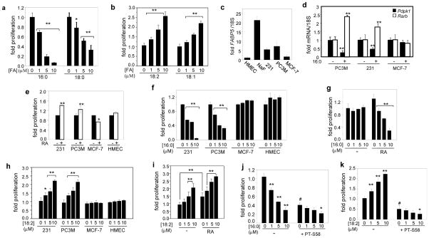
Figure 3. SLCFA inhibit and ULCFA induce carcinoma cell growth.
a), b) NaF cells were treated with 16:0, 18:0, 18:2, or 18:1 for 4 days. Proliferation was assessed by MTT assays. c) FABP5 mRNA in normal human mammary epithelial cells (HMEC), NaF, MDA-MB-231 and MCF-7 mammary carcinoma cells, and PC3M prostate carcinoma cells. d) Pdpk1 and Rarb mRNA in denoted cells treated with 16:0 (10 μM, 6 h.). e) Cells were treated with vehicle (−) or RA (1 μM) for 4 days and proliferation assessed by MTT assays. f) Effect of 16:0 (4 days) on proliferation of denoted cells. g) Cells were cultured in CT-FBS, treated with 16:0 in the absence or presence of RA (0.2 μM, 4 days), and proliferation assessed by MTT assays. **p<0.01, by unpaired t-test vs. cells cultured in CT-FBS replenished with RA. h) Effect of 18:2 (4 days) on proliferation of denoted cells assessed by MTT assays. i) Cells were cultured in CT-FBS, co-treated with 18:2 and RA (0.2 μM) for 4 days and proliferation assessed by MTT assays. j), k) Effect of 16:0 (j) or 18:2 (k) on proliferation of NaF cell in the presence or absence of the PPARβ/δ antagonist PT-S58 (5 μM). #p<0.01 vs. untreated control, *p<0.05 vs. PT-S58 treatment without LCFA. Data in all panels are Mean ±SD. *p<0.05, **p<0.01 vs. the respective control. p-values were calculated by unpaired t-test.

