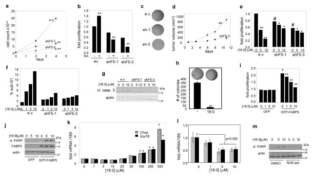
Figure 4. FABP5 mediates SLCFA-induced growth arrest and apoptosis.
a) Proliferation of cells expressing varying levels of FABP5 was assessed by cell counting. Mean±SD (n=4). **p<0.01, by unpaired t-test vs. e.v. expressing cells. b) MTT assays in cells treated with vehicle or RA (1 μM, 4 days). Mean±SD (n=4). **p<0.01, by unpaired t-test vs. untreated cells. c) Representative image (out of 3) of colonies formed in soft agar by NaF lines with varying expression levels of FABP5. See Fig. S5b for colony counts. d) NaF cells (2×10⁶) stably expressing empty vector or vector encoding FABP5shRNA (shF5-3, Fig. S2h) were transplanted into the mammary fat pad of NCr athymic female mice (n=5). Tumor growth was monitored. Mean ±SEM. e) MTT assays in NaF cells treated with 16:0 (4 days). Mean±SD (n=4). *p<0.05, **p<0.01 vs. untreated control, #p<0.01 vs. e.v. control. p-values were calculated by unpaired t-test. f) Cells were treated with 16:0 for 4 days and apoptosis evaluated by FACS analyses. Data from 1 out of 4 experiments are shown. g) Immunoblots of cleaved caspase 3 in NaF lines treated with 16:0 for 4 days. h) Representative image (out of 3) of colonies formed in soft agar by NaF cells treated with vehicle (−) or 16:0 (10 μM). **p<0.01 by unpaired t-test. i) MTT assays in MCF-7 cells overexpressing GFP-FABP5 or GFP control and treated with 16:0 (8 days). Mean±SD (n=4). *p<0.05, **p<0.01 vs. untreated control, #p<0.01 vs. e.v. control. j) Immunoblots of cleaved PARP in MCF-7 cells overexpressing GFP-FABP5 or GFP control and treated with 16:0 for 8 days. k) Levels of Chop and Grp78 mRNA in NaF cells treated with varying concentrations of 16:0 for 8 h. Mean ±SD (n=3). *p<0.05, *p<0.01 by unpaired t-test. l) Levels of Chop and Grp78 mRNA in NaF cells treated with 16:0 for 4 days. m) Immunoblots of cleaved PARP in NaF cells treated with 16:0 for 4 days in the presence or absence of antagonists for RARα (BMS19614), RARβ (LE135), or RARγ (MM11253) (1 μM each). Immunoblots in panels g, j, and m are representative of 3 independent experiments.

