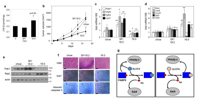
Figure 6. 16:0 shifts RA signalling and suppresses mammary tumor growth in vivo.
a) Concentrations of 16:0 in tumors of mice fed denoted diets. Mean±SD (n=6). **p<0.01 by unpaired t-test vs. chow control. b) NaF cells (2×10⁶) were transplanted into the mammary fat pad of NCr athymic female mice fed denoted diets and tumor growth was monitored. Mean±SD (n=6). **p<0.01 by unpaired t-test vs. chow control. c), d) mRNAs for PPARβ/δ (c) or RAR target genes (d) in mice fed denoted diets. Mean±SD (n=6). *p<0.05, **p<0.01 calculated by unpaired t-test. e) Immunoblots demonstrating expression of PDPK1 and RARβ in tumors of mice fed denoted diets. Blots are representative data from 3 individual mice per group. f) Immunohistochemistry analyses demonstrating expression of Ki67 and cleaved caspase 3 in tumors of mice fed denoted diets. Representative images out of three tumors per group are shown. Magnification 400x, scale bars represent 50 μm. g) A model illustrating the mechanism by which SLCFA and ULCFA regulate RA signaling. Left: SLCFA compete with RA and other PPARβ/δ ligands for binding FABP5, divert RA to RAR and inhibit PPARβ/δ activation. Right: ULCFA bind to FABP5 and divert RA to RAR. ULCFA are delivered to PPARβ/δ by FABP5 and activate it.

