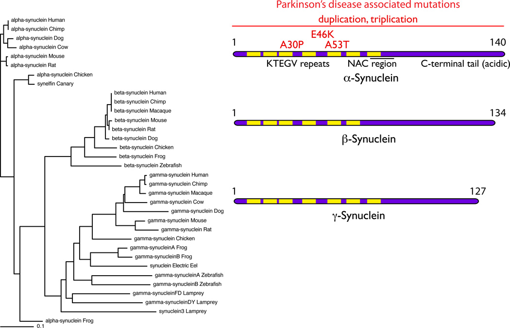
Figure 2. The Synuclein family.
The tree on the left of the figure shows that there are several synuclein homologues in many vertebrate species. These separate into three groups, identified as α-, β- and γ- synuclein and, interestingly, most species have one of each homologue. Exceptions to this general rule include species such as the lamprey and zebrafish, which appear to have evolved multiple γ- synuclein homologues but lack an α-synuclein homologue. On the right are ideograms of the proteins. The characteristic KTEGV repeats, the number of which vary between homologues are indicated in yellow. Mutations in α-synuclein, shown in red, are associated with autosomal dominant Parkinson’s disease and include three point mutations and multiplications of the whole SNCA locus (indicated by the horizontal line).

