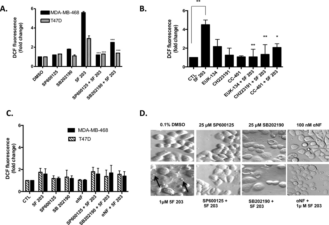
Figure 4.
JNK and p38 contribute to 5F 203-mediated increases in ROS production, and 5F 203-mediated apoptotic body formation, at least partially, relies on AhR, p38, and JNK signaling in breast cancer cells. (A) MDA-MB-468 and T47D cells were pretreated for 1 h with SP600125 or SB202190 before being exposed to 1 µM 5F 203 for 6 h. Otherwise, cells were exposed to medium containing 0.1% DMS0, 5F 203, or the respective inhibitors for 6 h. ***p < 0.001 compared to treatment with 5F 203 alone. (B) MDA-MB-468 cells were pretreated for 1 h with CC-401, CH223191, or EUK-134 before exposure to 1 µM 5F 203 for 6 h. Otherwise, cells were exposed to medium containing 0.1% DMS0, 5F 203, or the respective inhibitors for 6 h. *p < 0.05 or **p < 0.01 compared to treatment with 5F 203 alone. (C) MDA-MB-468 and T47D cells were pretreated for 30 min with SP600125 or SB202190 followed by 1 or 3 h of exposure to 5F 203, respectively. Otherwise, cells were exposed to medium containing 0.1% DMS0, 5F 203, or the respective inhibitors for either 1 or 3 h. ROS levels were evaluated using flow cytometry. (D) MDA-MB-468 breast cancer cells were exposed to 5F 203 (6 h) with or without pretreatments with 100 nM αNF, 25 µM p38 SB202190, or 25 µM SP600125 (1 h) before cells were visualized by relief contrast microscopy. Magnification, 200×. Apoptotic bodies are indicated by arrows.

