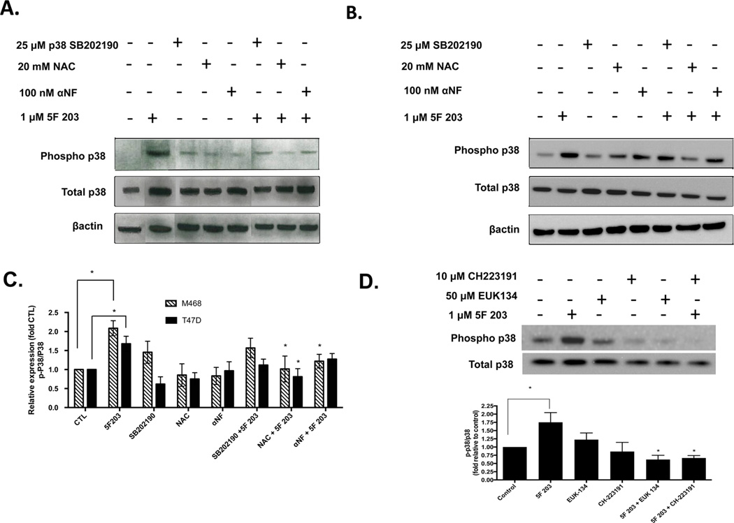
Figure 6.
AhR antagonists suppress 5F 203-mediated p38 phosphorylation in breast cancer cells. (A) MDA-MB-468 and (B) T47D cells were treated with 0.1% DMSO, αNF, NAC, SB202190, or 1 µM 5F 203 for 6 h or were pretreated for 1 h with αNF (100 nM), NAC (20 mM), or SB202190 (25 µM) before being exposed to 1 µM 5F 203 for 6 h. Following treatment, cells were lysed, and protein content was determined and resolved using western blotting as described in Materials and Methods. Gel is of a representative blot, and sections within the blot were rearranged based on treatments specified. (C) Data is representative of three independent experiments. Graphs based on arbitrary units of ratios densitometry determinations for phospho-proteins relative to total protein and used to quantify the extent of activation. (D) MDA-MB-468 cells were exposed to 5F 203 (6 h) with or without EUK-134 or CH223191 pretreatment, and western blotting analysis was performed along with densitometry readings. Data represent the mean of at least three independent experiments; bars, SEM. Except where indicated by lines, *p < 0.05 compared to cells exposed to 5F 203 only.

