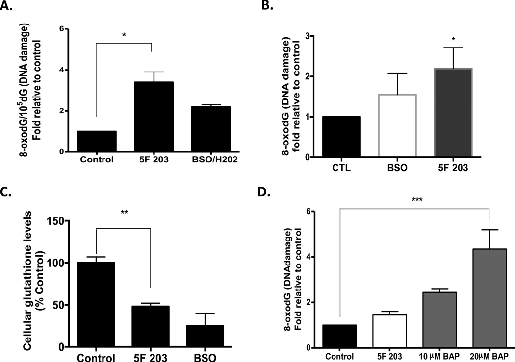
Figure 8.
5F 203 induces oxidative DNA damage in MDA-MB-468 breast cancer but not MCF-10A breast epithelial cells. (A) Oxidative DNA damage was determined by measuring the levels of 8-oxodG/105 dG. MDA-MB-468 cells were treated with 1 µM 5F 203 for 12 h or pretreated for 18 h with 100 µM BSO followed by the addition of 500 µM H2O2 in the medium for 30 min. Data represent the mean ± SEM of at least three independent experiments. *p < 0.05. (B) Oxidative DNA damage was also determined using the OxyDNA assay with the treatments described in (A). Data represent the mean ± SEM of at least three independent experiments performed in duplicate. *p < 0.05 compared to control. (C) Cells were exposed to medium containing 0.1% DMSO (negative control, 12h), 1 µM 5F 203 (12 h), or buthionine sulfoxide (BSO, 100 mM, 18 h) before the glutathione assay was performed. **p < 0.01. (D) MCF-10A breast epithelial cells were exposed to 0.1% DMSO, 5F 203 (1 µM), or benzo[a]pyrene (10 and 20 µM) for 24 h as outlined in Materials and Methods. Data represent the mean ± SEM of at least three independent experiments. ***p < 0.001.

