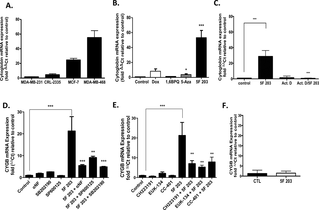
Figure 9.
5F 203 induces cytoglobin expression in sensitive breast cancer cells, which depends, in part, on AhR, p38, or JNK signaling. (A) MDA-MB-468, MCF-7, CRL2335, and MDA-MB-231 cells were exposed for 24 h to medium containing 0.1% DMSO or 1 µM 5F 203. RNA was extracted, and gene expression was evaluated using real-time PCR as outlined in Materials and Methods. Data represent the mean ± SEM of at least three independent experiments. (B) MDA-MB-468 breast cancer cells were exposed to medium containing 0.1% DMSO, 1 µM 5F 203, 10 µM 5-aza-2′-deoxycytidine (5-Aza), 1 µM doxorubicin (Dox), or 5 µM 1,6-BPQ for 24 h. RNA was extracted from cells, and cytoglobin mRNA expression was determined in accordance with Materials and Methods. *p < 0.05 and ***p < 0.001 compared to control. Data represent the mean ± SEM of at least three independent experiments performed in triplicate. (C) MDA-MB-468 breast cancer cells were exposed to medium containing 0.1% DMSO, 250 nM 5F 203, 5 µM actinomycin D (Act. D), or 5F 203 in combination with Act. D for 12 h before cells were harvested, RNA was extracted, and mRNA expression was determined as outlined in Materials and Methods. Data represent the mean ± SEM of at least three independent experiments. ***p < 0.001 as designated by line comparison. Otherwise, **p < 0.01 or ***p <0.001 compared to cells exposed to 5F 203 only. (D) MDA-MB-468 breast cancer cells were exposed for 12 h to medium containing 0.1% DMSO, 100 nM αNF, 25 µM SP600125, 25 µM p38 SB202190, 1 µM 5F 203 in the presence or absence of inhibitor pretreatments before gene expression was evaluated using quantitative-PCR in accordance with Materials and Methods. ***p < 0.001 as designated by line comparison. Otherwise, **p < 0.01 or ***p < 0.001 compared to cells exposed to 5F 203 only. Data represent the mean ± SEM of three independent experiments. (E) MDA-MB-468 cancer cells were exposed for 12 h to medium containing 0.1% DMSO, 10 µM CH223191, 10 µM CC-401, 50 µM EUK-134, or 1 µM 5F 203 in the presence or absence of inhibitor or antioxidant pretreatments before gene expression was evaluated using quantitative-PCR in accordance with Materials and Methods. ***p < 0.01 as designated by line comparison. Otherwise, **p < 0.01 compared to cells exposed to 5F 203 only. (F) AHR100 cells were exposed to 0.1% DMSO or 1 µM 5F 203 for 12 h before undergoing cytoglobin mRNA expression analysis using quantitative PCR analysis. Data represent the mean ± SEM of three independent experiments.

