
Figure 3.
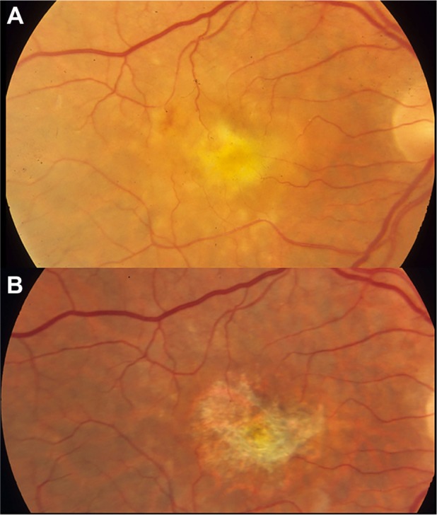
An eye with neovascularization-associated RPE atrophy at the baseline (A) and 30 months after anti-VEGF treatment (B).
Notes: With resolution of subretinal fluid, fibrin, and thin fibrovascular material at the baseline, the eye has developed marked RPE loss in the central macula.
Abbreviations: RPE, retinal pigment epithelium; VEGF, vascular endothelial growth factor.