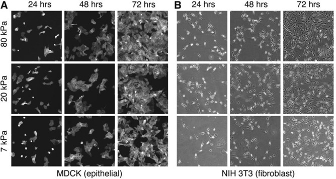
Figure 5. Biocompatibility.
Fluorescence and phase contrast images showing the proliferation of (A) MDCK epithelial and (B) NIH/3T3 fibroblast cells at 24, 48, and 72 hours on substrates formed by soft PDMS elastomers with storage moduli of 7kPa (SEL), 20kPa (SE5), and 80kPa (SE30). Images are 750µm×750µm.

