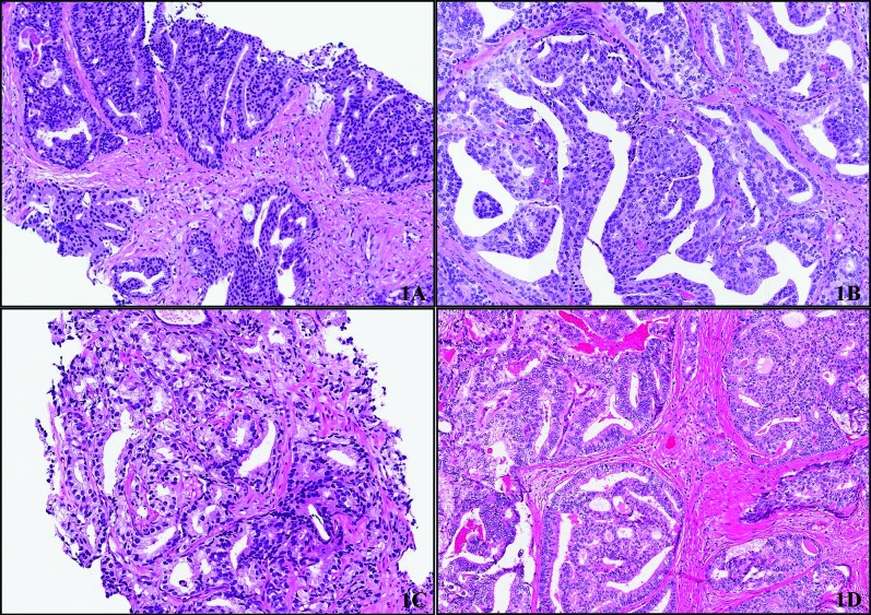
Fig. 1.
(A) Example of consensus diagnosis for prostatic ductal adenocarcinoma on transrectal ultrasound (TRUS)-guided biopsy. Cribriform structures are lined by tall columnar cells with amphophilic cytoplasm and high grade nuclei. (B) Corresponding RP to biopsy in 1A showing similar morphologic features, including papillary and cribriform structures. (C) Conventional acinar prostatic carcinoma on TRUS guided biopsy (Gleason score 4+3 =7) showing gland fusion and irregular lumina. There are no features that are characteristic of prostatic ductal adenocarcinoma. (D) Corresponding RP to biopsy shown in 1C showing features of prostatic ducal adenocarcinoma including large glands lined by tall columnar cells with amphophilic cytoplasm and high grade nuclei. Cells are arranged in cribriform structures with slit-like spaces.

