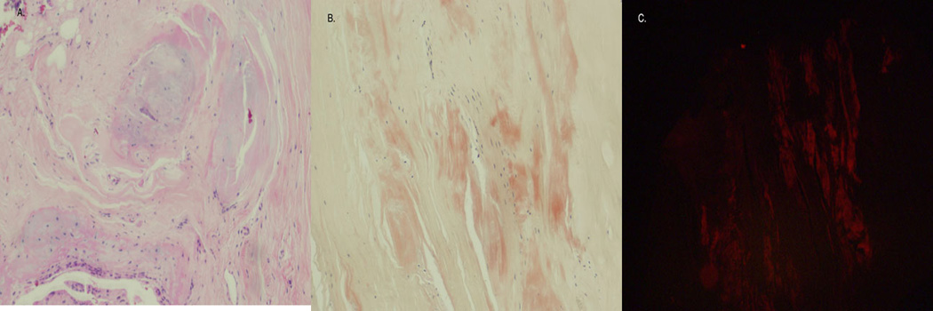
Figure 2. Amyloid deposition in the bone marrow (bone marrow biopsy).

A. the bone marrow biopsy exhibited normal hematopoietic cells (not shown) with extensive fibrous tissue present adjacent to the marrow. (H&E 200×). B. Congo Red stain shows multiple amorphous orange-red deposits within the fibrous tissue. (H&E 200×). C. Polarized light microscopy confirmed amyloid deposition in the fibrous tissue.
