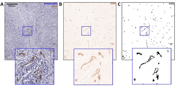
Figure 1. Immunostained blood vessels are automatically segmented in whole-slide images.

In this figure, image tiles of 1600×1600 px are shown and an image detail is enlarged. A. Original image region, B. result of color deconvolution, C. result of thresholding and morphological post-processing.
